Age-related macular degeneration (AMD): what matters
Age-related macular degeneration is the most common cause of central vision loss after 60. It almost never causes total blindness — but it affects precisely the functions used all day. Understanding the difference between its two forms is what determines how urgently to act.
What the macula is, and why it matters so much
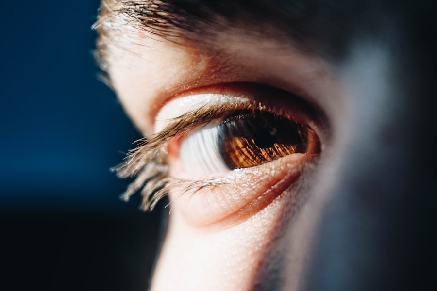
The macula is a small area at the centre of the retina, only about five millimetres across. It holds the highest density of photoreceptors in the eye and is responsible for everything that requires sharp vision: reading, recognising faces, driving, close work, telling colours apart.
Age-related macular degeneration (AMD) is a process in which the macula is gradually damaged with age. Peripheral vision usually remains intact — which is why the disease almost never causes total blindness — but the functions a person uses all day are precisely the ones affected.
Two forms, two different stories
The dry form
This is the common one — roughly nine cases in ten. It develops slowly, sometimes over years. Deposits called drusen accumulate beneath the macula, and later the cells themselves thin out. In its early stages it may be barely noticeable, and is often found on a routine examination.
The wet form
Far less common, but responsible for most cases of rapid vision loss. Here, new and abnormal blood vessels grow beneath the retina. They leak fluid and blood, the macula swells, and vision can change over days to weeks.
The distinction is not academic: in the wet form, how quickly you act changes the outcome.
Signs worth knowing
- Straight lines that look bent — a door frame, floor tiles, lines of text. This is the most characteristic sign, and usually the first.
- A blurred or missing patch in the centre — while the periphery stays sharp.
- Needing more light to read than before, and slow recovery moving from dark into light.
- Difficulty recognising faces at a distance that used to be comfortable.
- Colours looking faded.
Here too, the healthy eye compensates. Many patients discover the condition by accident — while covering one eye for some entirely unrelated reason.
An Amsler grid is a squared chart with a dot at its centre. Wearing your reading glasses, cover one eye, focus on the central dot, and check whether every line looks straight and continuous.
Wavy, blurred or missing lines — and above all a change since yesterday — call for an examination soon. It does not replace a doctor, but it is the simplest way to catch a change early.
How it is diagnosed
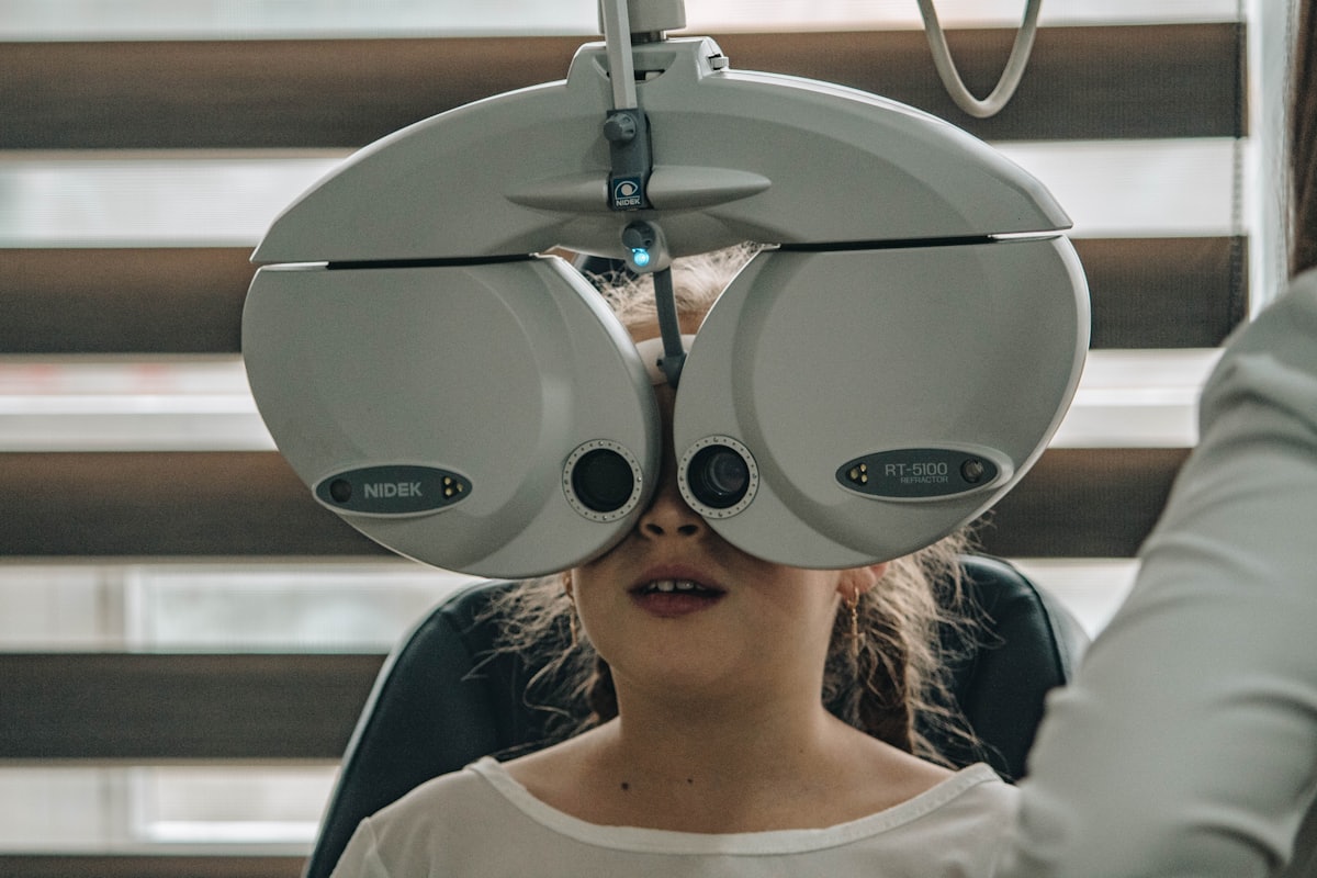
Diagnosis rests on a dilated examination of the retina, together with imaging:
- OCT — a non-invasive, painless optical scan showing a cross-section of the macular layers to within microns. It reveals fluid, swelling and drusen, and it is also the tool used to follow the response to treatment.
- Fundus photography — a record for comparison over time.
- Angiography — a study that shows the retinal blood vessels and helps locate leakage where the wet form is suspected.
What treatment involves today
For the wet form
The standard treatment is intravitreal injections of anti-VEGF drugs, which block the factor driving the growth of abnormal vessels. The injection is given under sterile conditions after anaesthetic drops and takes minutes.
It is given as an initial series, then at intervals set by the response and by the OCT findings. Two things are worth knowing in advance: treatment usually stabilises vision and sometimes improves it, but does not cure the disease; and regularity matters — a long gap can allow the leakage to return.
For the dry form
There is at present no treatment that restores cells already lost. What does exist, and is well established:
- AREDS2-formulation supplements — shown to reduce the risk of progression in particular groups of patients with intermediate disease. They are not right for everyone, and they are not a medicine — the decision should be made with a doctor.
- Regular monitoring — to catch a conversion to the wet form at a stage where it can be treated.
- In recent years, new treatments for advanced dry disease have been approved in some countries, and this is a field that is changing. It is worth asking what is relevant to your specific situation.
What is genuinely in your control
Age and genetics cannot be changed. Two things can:
- Smoking — by a wide margin the most significant modifiable risk factor for macular degeneration. Stopping reduces the risk even later in life.
- Blood pressure, lipids and diet — a diet rich in leafy vegetables and fish, and general metabolic control, are associated with lower risk.
UV-protective sunglasses are a sensible recommendation in any case, even though the direct link to macular degeneration is not clear-cut.
Sudden distortion of straight lines, a new patch in the centre of vision, or a rapid drop in vision — particularly in someone already diagnosed with macular degeneration — may indicate a conversion to the wet form. That warrants an examination within days, not weeks.
Living with the condition
Macular degeneration does not mean blindness. Peripheral vision remains, and walking, orientation and independence are preserved in most cases. What it does require is adaptation: better lighting, magnifiers, high-contrast text, and sometimes low-vision services. For someone whose central vision has been affected, these adjustments make a large difference to everyday function.
The information in this article is general information only. It does not constitute medical advice or a diagnosis, and it is not a substitute for examination by a doctor. Every case is different, and nothing here should be relied on for medical decisions. In the event of a sudden loss of vision, flashes of light, a sudden shower of floaters or a shadow falling across your vision, seek urgent medical care immediately.