Retinal detachment: warning signs you should never ignore
Retinal detachment is one of the few conditions in ophthalmology where the difference between coming in today and coming in next week is the difference between vision preserved and permanent damage. The warning signs almost always appear — the difficulty is how easy they are to dismiss.

What the retina is, and what happens when it detaches
The retina is a very thin layer of tissue — less than a millimetre thick — lining the inside of the back of the eye. It is what captures light and converts it into the nerve signals sent to the brain. Without it, the eye is a lens with no picture.
Beneath the retina sits a layer of cells that supplies it with oxygen and nutrients. A retinal detachment is a condition in which the retina physically separates from that layer. From the moment it separates, the supply stops and the light-sensitive cells begin to suffer. The longer this lasts, the more of the damage becomes permanent.
In most cases the process begins with a small tear in the retina. Fluid from the vitreous cavity — the clear gel that fills the eye — passes through the tear and starts lifting the retina off the wall, rather like wallpaper peeling once water gets behind it. This is why treating a tear before a detachment forms is incomparably simpler than treating the detachment itself.
The four warning signs
- Flashes of light — brief flashes, usually at the edge of the visual field, most noticeable in a dark room or on rapid eye movement. They are caused by the vitreous pulling mechanically on the retina.
- A sudden shower of floaters — specks, threads or cobwebs drifting across your vision. The occasional floater is common and usually harmless; a sudden shower of them, or a new large dark floater, is a different matter.
- A shadow, curtain or dark wall — a black or grey area covering part of the visual field, often from above or from the side, and advancing over hours. This already indicates an active detachment.
- A sudden drop in acuity — particularly when it affects the centre of vision.
Two things are worth knowing in advance. First: a retinal detachment is almost always painless. The absence of pain is not reassuring and rules nothing out. Second: the other eye compensates very well, so it is possible to lose a substantial part of the visual field in one eye without noticing — until you close the good one. If something feels different, simply cover each eye in turn and compare.
If any of these signs has appeared in the last few days, do not wait for an appointment. Call the clinic, and if there is no immediate answer, go to an eye emergency room the same day.
A retinal tear treated in time is usually dealt with by a short laser treatment in the clinic. The same tear a week later may already require surgery.
Why every hour counts
The centre of the retina is called the macula, and it is responsible for sharp vision — reading, recognising faces, driving. As long as the detachment has not reached the macula, the chance of good vision after successful surgery is significantly higher. Once the macula detaches, functional recovery is less complete, and every additional day lowers the ceiling.
This is why, in practice, retinal detachment is treated as two quite different levels of urgency — before the macula is involved, and after. The difference between them is sometimes measured in hours.
Who is at higher risk
- High myopia (short-sightedness) — a longer eye, and a thinner, more stretched retina. This is the most significant risk factor at younger ages.
- Age 50 to 75 — the years in which the vitreous naturally shrinks and separates from the retina, a process that can pull and create a tear.
- After cataract surgery — the risk rises, particularly in the first years afterwards.
- Eye trauma — including injuries that seemed minor, and including years later.
- A previous detachment — in the same eye or the other one, or a family history.
- Known retinal disease — including advanced diabetic retinopathy and inherited retinal conditions.
Being in one of these groups is no reason to live on alert, but it is a good reason to know the four signs and to know who to call.
What the examination involves
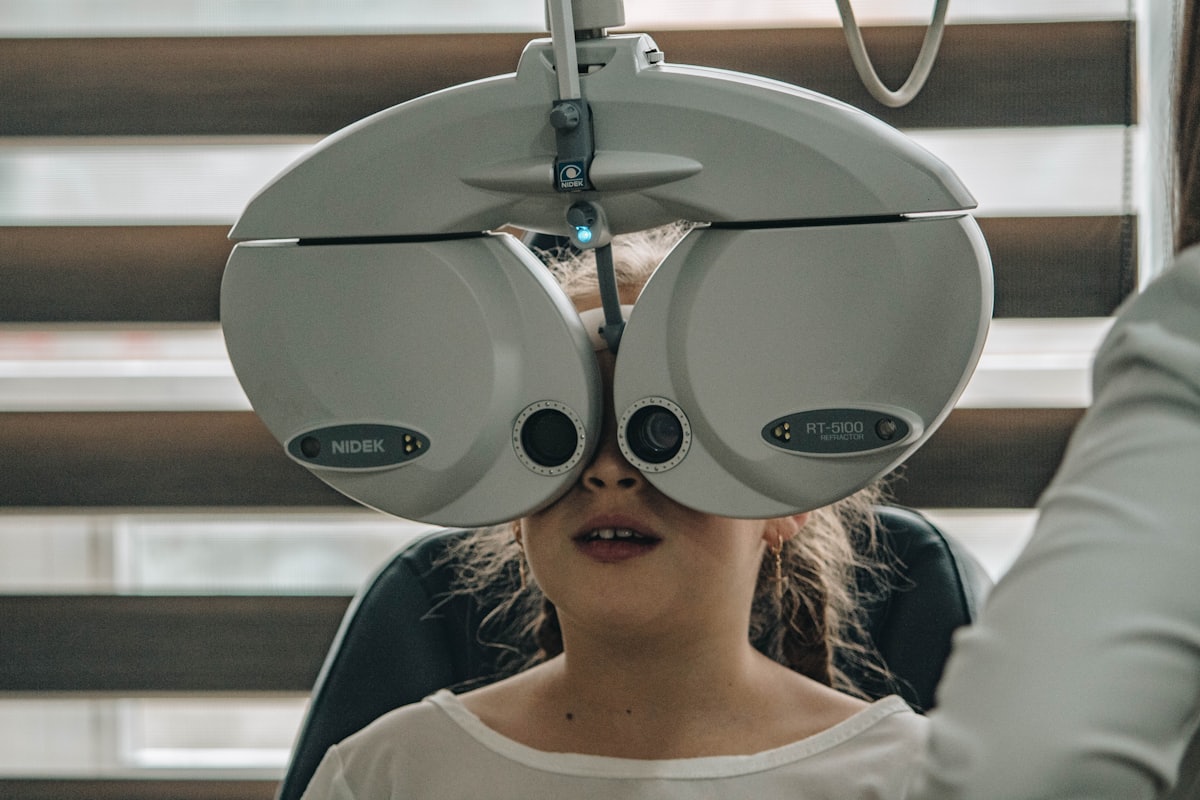
The central test is a dilated fundus examination. Drops are used to widen the pupil, and after around 20 minutes the whole retina can be examined — including the far periphery, where most tears occur and which cannot be seen without dilation.
Depending on the findings, an OCT scan is also used: a non-invasive optical scan showing a cross-section through the retinal layers, which establishes whether the macula is involved. Where bleeding into the vitreous obscures the view, an ultrasound of the eye shows what lies behind it.
Please note: after dilation vision is blurred for several hours and you should not drive. It is best to come with someone, and to bring sunglasses.
Treatment options
A retinal tear without detachment
Usually treated with laser (retinopexy) or freezing. The treatment is done in the clinic, takes minutes, and creates a seal around the tear that stops fluid getting through. It is preventive — and it is the main reason to come early.
An established detachment
Requires surgery. There are three main approaches, and the choice between them depends on where the detachment is, how large it is, the state of the vitreous, and age:
- Pneumatic retinopexy — a gas bubble, suitable for some upper detachments.
- Scleral buckle — a silicone band placed around the eye to bring the wall back to the retina.
- Vitrectomy — removing the vitreous and replacing it with gas or silicone oil, which holds the retina in place until it reattaches.
After surgery with gas, a specific head position is sometimes required for a few days, along with avoiding air travel until the gas has absorbed. The details vary from operation to operation and are explained in advance.
In most cases the retina is successfully reattached with a single operation, and in some a further procedure is needed. It is worth separating two different questions: whether the retina was reattached — and what the vision will be. The second depends mainly on whether the macula detached, and for how long.
Second opinions
An active detachment is not a situation in which to wait for a second opinion — there, time is the deciding factor. Before planned surgery, however, when the diagnosis is not clear-cut, or when there is disagreement about which operation is appropriate, a further opinion is an entirely reasonable step, and the clinic takes such enquiries regularly.
The information in this article is general information only. It does not constitute medical advice or a diagnosis, and it is not a substitute for examination by a doctor. Every case is different, and nothing here should be relied on for medical decisions. In the event of a sudden loss of vision, flashes of light, a sudden shower of floaters or a shadow falling across your vision, seek urgent medical care immediately.