Uveitis: a short guide for patients
Uveitis is inflammation of the middle layer of the wall of the eye. It can look like an innocent red eye, and sometimes cannot be seen at all — yet it remains one of the significant causes of preventable vision loss.
What the uvea is, and what uveitis means
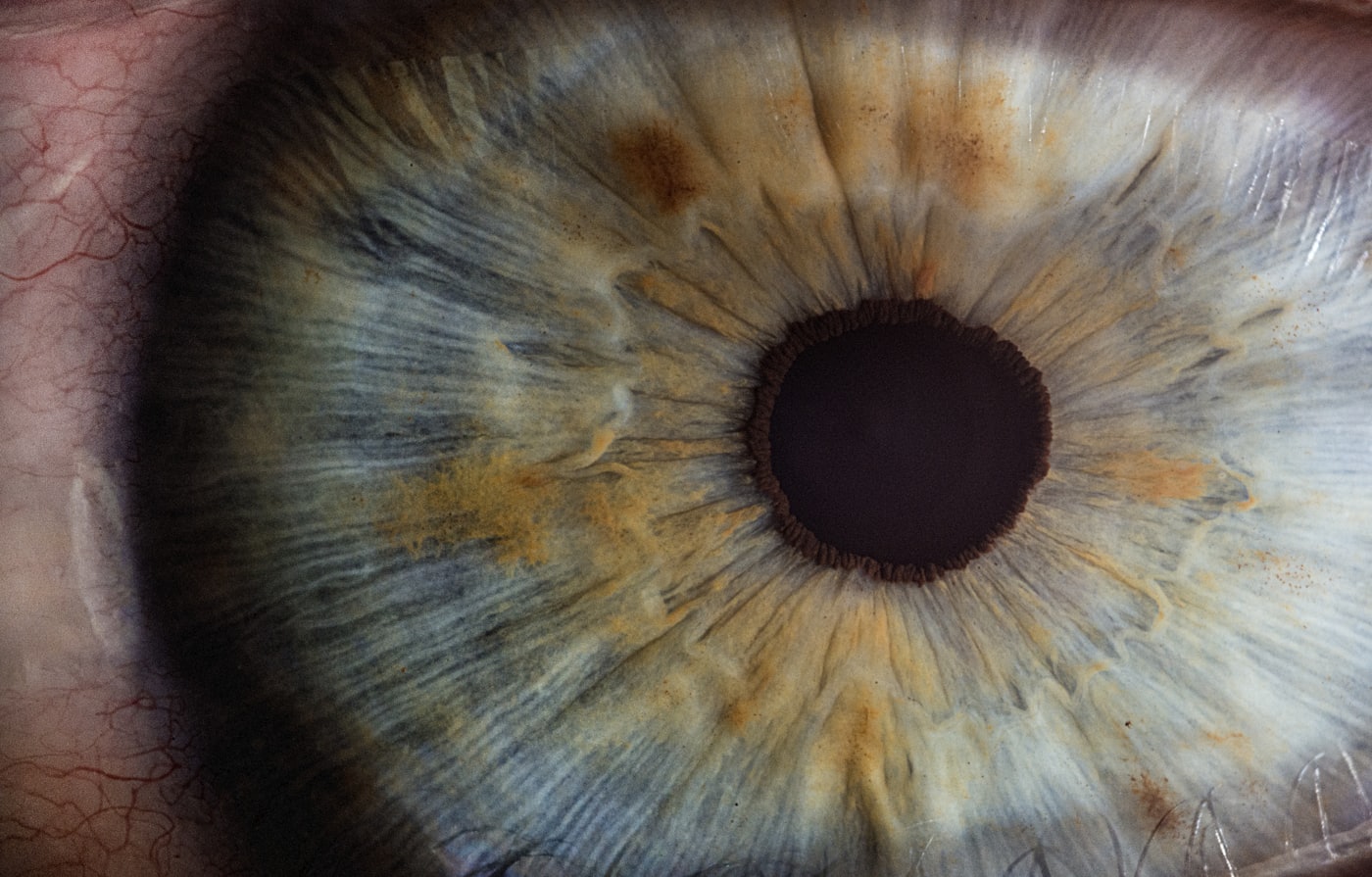
The uvea is the middle, vascular layer of the wall of the eye. It has three parts: the iris — the coloured part you can see from outside; the ciliary body behind it, which produces the fluid inside the eye and controls focusing; and the choroid, the layer of blood vessels that nourishes the retina from behind.
Uveitis is inflammation of that layer. It is not a single disease but a group of conditions sharing an inflammatory process inside the eye — and differing widely in cause, course and treatment.
It is classified by where the inflammation sits:
- Anterior uveitis — the most common. The inflammation centres on the iris and ciliary body, and usually presents with redness and pain.
- Intermediate uveitis — in the vitreous cavity, often without redness or pain, with floaters as the main complaint.
- Posterior uveitis — involving the choroid and the retina. This is the form in which a retina specialist's involvement matters most.
- Panuveitis — inflammation throughout the uvea.
Symptoms
The picture varies with the location of the inflammation, and that is exactly what makes it misleading:
- Redness, usually concentrated around the cornea rather than across the whole white of the eye.
- A dull ache or pressure in the eye.
- Marked sensitivity to light (photophobia) — very characteristic of the anterior form.
- Blurred vision or a sense of haze.
- Floaters and specks in the visual field.
- Watering, and a small or irregular pupil.
One important point: the posterior and intermediate forms can be entirely quiet — no redness, no pain — showing up only as blurring or floaters. A white, comfortable eye does not rule out active inflammation deeper inside.
A red eye with pain and marked light sensitivity, or with reduced vision, is not ordinary conjunctivitis and is not something to wait out. It needs to be examined by an ophthalmologist, ideally the same day or the next.
This is especially true for anyone previously diagnosed with uveitis, or with a known autoimmune condition — in whom these symptoms usually represent a flare.
What causes it
In roughly half of cases no identifiable cause is found and the condition is termed idiopathic. In the rest, the main causes are:
- Systemic autoimmune and inflammatory disease — among others, conditions associated with the genetic marker HLA-B27 (such as ankylosing spondylitis and inflammatory bowel disease), sarcoidosis, Behçet's disease, and juvenile idiopathic arthritis.
- Infection — herpes viruses, toxoplasmosis, tuberculosis, syphilis and others.
- Trauma or eye surgery.
- Rarely, a reaction to particular medications.
Diagnosing uveitis therefore does not stop at the eye. In some patients the eye is the first sign of a systemic disease not yet diagnosed, and the right work-up can change treatment well beyond ophthalmology.
Why it should not be left
Active inflammation left untreated can leave cumulative damage, even when it is tolerable:
- Macular oedema — the most common cause of permanent vision loss in uveitis.
- Adhesions between the iris and the lens, distorting the pupil.
- Raised intraocular pressure and glaucoma — from the inflammation itself and sometimes from steroid treatment.
- Early cataract.
- In posterior forms — retinal scarring, and in some cases retinal detachment.
The logic of treatment is to put the inflammation out quickly and keep it out — not merely to ease the symptom.
How it is diagnosed
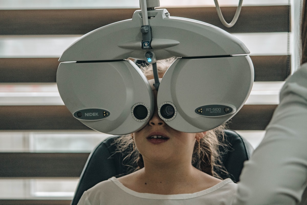
The basis is a slit-lamp examination, in which inflammatory cells can be seen floating in the fluid inside the eye, together with measurement of intraocular pressure and a dilated examination of the retina.
Depending on the findings, this is supplemented by:
- OCT of the macula — to detect swelling, the complication that matters most for vision.
- Angiography — to assess involvement of the retinal vessels.
- Blood tests and imaging — to look for a systemic or infectious cause, guided by the clinical picture. Not every patient needs a broad work-up, and testing in every direction is not better.
In many cases treatment is shared with a rheumatologist or physician, particularly where systemic therapy is required.
Treatment
Treatment is determined by the location of the inflammation, its severity and its cause. The main tools:
- Steroid drops — the backbone of treatment in the anterior form, tapered and monitored.
- Dilating drops — which relieve pain and prevent adhesions.
- Steroid injections around or into the eye — for posterior forms and for macular oedema.
- Systemic treatment — oral steroids, and in chronic cases immunosuppressants or biologic agents that allow steroids to be withdrawn.
- Targeted anti-infective treatment where the cause is infectious — here a steroid alone can make matters worse, which is why the diagnosis comes before the treatment.
Uveitis tends to recur. Structured follow-up, including during quiet periods, is part of the treatment rather than an addition to it — not least to monitor intraocular pressure and macular oedema before either is noticeable.
The information in this article is general information only. It does not constitute medical advice or a diagnosis, and it is not a substitute for examination by a doctor. Every case is different, and nothing here should be relied on for medical decisions. In the event of a sudden loss of vision, flashes of light, a sudden shower of floaters or a shadow falling across your vision, seek urgent medical care immediately.